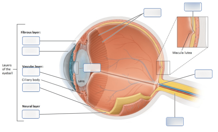
Art-labeling activity: sagittal section of internal structures of the eye – Embark on an art-labeling adventure with the sagittal section of the eye, where intricate internal structures unfold before your eyes. This engaging activity invites you to explore the depths of human anatomy, transforming complex structures into a canvas for discovery.
Through the lens of art, you’ll uncover the intricacies of the eye, identifying key components and gaining a deeper understanding of their functions. Prepare to immerse yourself in a world of anatomical exploration and artistic expression.
Sagittal Section of Internal Structures of the Eye

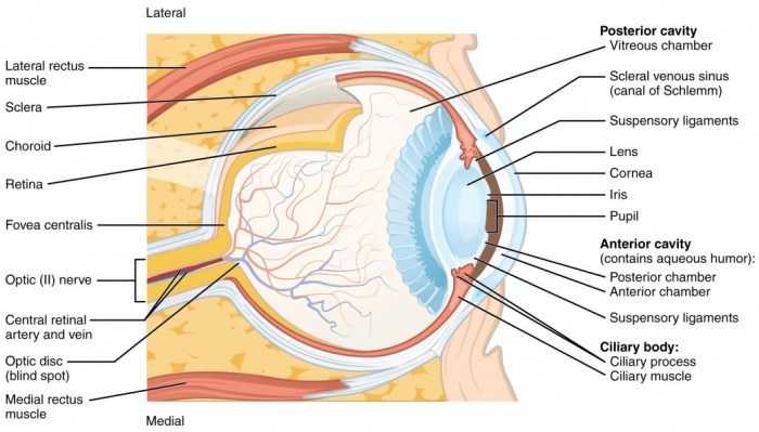
The sagittal section of the eye provides a detailed view of the internal structures of the eye, as seen in a plane that divides the eye into left and right halves. This section allows visualization of the eye’s anatomy from the cornea to the retina.
Visible Internal Structures, Art-labeling activity: sagittal section of internal structures of the eye
Within the sagittal section, several key internal structures are visible:
- Cornea:The clear, outermost layer of the eye that refracts light.
- Lens:A transparent structure that further focuses light onto the retina.
- Pupil:The black opening in the center of the iris that allows light to enter the eye.
- Iris:The colored part of the eye that controls the size of the pupil.
- Retina:The light-sensitive layer at the back of the eye that converts light into electrical signals.
- Vitreous Humor:A gel-like substance that fills the main chamber of the eye behind the lens.
- Optic Nerve:The bundle of nerve fibers that transmits visual information from the retina to the brain.
Labeling Activity

Instructions:
- Examine the sagittal section of the eye provided.
- Identify the internal structures listed in the table below.
- Label each structure directly on the section using the corresponding number.
Table of Labels:
| Number | Label | Description |
|---|---|---|
| 1 | Cornea | Clear, outermost layer of the eye |
| 2 | Lens | Transparent structure that focuses light |
| 3 | Pupil | Black opening in the iris |
| 4 | Iris | Colored part of the eye |
| 5 | Retina | Light-sensitive layer at the back of the eye |
| 6 | Vitreous Humor | Gel-like substance filling the main chamber |
| 7 | Optic Nerve | Nerve fibers transmitting visual information |
Educational Applications: Art-labeling Activity: Sagittal Section Of Internal Structures Of The Eye

Art-labeling activities are valuable in anatomy education as they:
- Enhance understanding:Labeling forces students to actively engage with the anatomical structures, improving their comprehension and retention.
- Promote observation skills:Students must carefully observe the structures to accurately label them, developing their visual analysis abilities.
- Facilitate discussion:Labeling activities can be used as a starting point for discussions about the relationships and functions of different anatomical structures.
Integration into Lesson Plans:
- Use labeling activities as a pre-lab exercise to prepare students for dissections or simulations.
- Incorporate labeling into lectures to reinforce anatomical concepts and provide visual aids.
- Assign labeling activities as homework or online assignments to encourage independent learning.
Art-Labeling Techniques

Effective art-labeling activities involve:
- Clear and accurate labels:Labels should be concise, descriptive, and placed near the corresponding structures.
- Visual aids:Use colors, arrows, or highlighting to draw attention to specific features.
- Multiple modalities:Combine digital drawing, painting, or collage to enhance engagement and cater to different learning styles.
- Peer review:Encourage students to review each other’s labels to foster collaboration and improve accuracy.
Frequently Asked Questions
What is the purpose of art-labeling activities in anatomy?
Art-labeling activities in anatomy provide a unique and engaging way to enhance students’ understanding of anatomical structures. By actively labeling and identifying components, students develop a deeper comprehension and retention of anatomical knowledge.
How can art-labeling activities be integrated into lesson plans?
Art-labeling activities can be seamlessly integrated into lesson plans by providing students with diagrams or images of anatomical structures. Students can then engage in the labeling process, using different art techniques to enhance their understanding and retention of the material.